


Human iPSC-derived Retinal pigment epithelium cells(iRPE)
Product Overview
The Retinal Pigment Epithelium (RPE) is a layer of hexagonally arranged, pigmented cells located between the neural retina and the choroid. RPE cells play a crucial role in maintaining the visual system by providing essential nutrients to photoreceptors, phagocytosing the spent outer segments of these photoreceptors, facilitating the visual/retinoid cycle to produce photosensitive derivatives of vitamin A, absorbing stray light to enhance vision, and secreting vascular endothelial growth factor (VEGF) to support the choroidal blood vessels. Abnormalities in the RPE can lead to a wide range of retinal degenerative diseases, notably age-related macular degeneration (AMD), which can result in severe vision loss. Currently, medication options for AMD are limited, prompting increasing interest in RPE cell transplantation as a potential treatment strategy.
To support researchers in understanding basic ocular biology and developing treatments for ocular diseases, iRPE cells represent a highly pure population of research-grade RPE cells derived from human iPSCs. These cells have been offering by iCells International, enable a wide range of applications spanning disease research, drug discovery, safety and toxicity testing, and regenerative medicine. Available in commercial quantities, iRPE possess the following features:
Mature around 2 weeks post-thaw
Cell purity greater than 95%
Cobblestone morphology growth
Abundant melanin deposition
Phagocytotic activity
Polarized secretion function
Biological Source | Human iPSCs |
Disease | Normal |
Growth Mode | Adherent |
Morphology | Hexagon, Pebble-like |
Number of cells | 1×10⁶cells/vial |
Product format | Frozen |
Storage | Vapor phase of liquid nitrogen |
Quality Control | Negative for HIV-1, HBV, HCV,CMV, mycoplasma, bacteria, yeast, and fungi |
Application | Research use only |
Performance Data
1)Cell morphology
iRPE is derived from the induced pluripotent stem cells (iPSC) and obtained high-purity RPE cells through a 45-day directed differentiation protocol in vitro.

Figure 1: Morphological map of cells at different stages of induced hiPSC differentiation into RPE in vitro
2)Cell purity
Cell purity greater than 95%

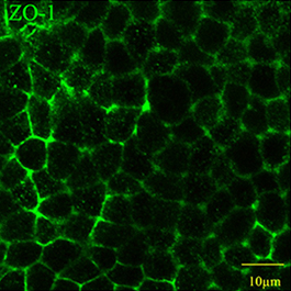
Figure 2: Immunostaining shows the expression of the RPE markers, ZO-1、MITF、RPE65、BEST1、CD140b and PMEL17

Figure 3: Flow cytometry shows the expression of the RPE markers, MITF、PMEL17、CD140b、BEST1
3)Cell function
iRPE exhibit phagocytotic activity as shown by pHrodo E.Coli Bioparticles conjugate for phagocytosis.

Figure 4: Phagocytosis activity
ZO-1 immunostaining of tight junctions illuminates the formation of an organized monolayer of polygonal RPE cells.

Figure 5: Immunostaining shows iRPE expressed ZO-1
iRPE has the function of polar secretion of PEDF and VEGF-A.

Figure 6: The significant difference in the concentrations of PEDF and VEGF-A in the apical and basal layer of the medium secreted by RPE grown in transwell